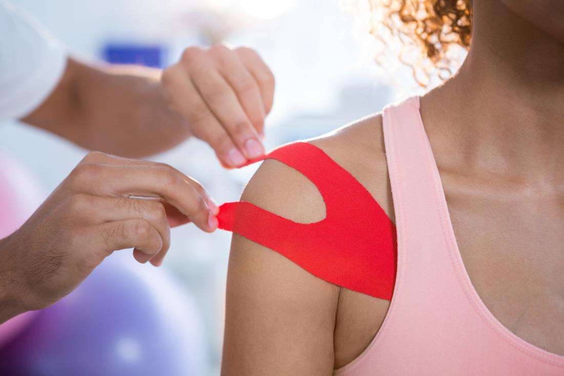
The experience of a shoulder dislocation is often sudden, deeply traumatic, and immediately incapacitating. Unlike a simple fracture or sprain, which might allow for limited function, a fully dislocated shoulder—where the humerus is forced entirely out of the glenoid socket—instantly renders the arm useless, accompanied by an intense, visceral pain. The shoulder joint, known anatomically as the glenohumeral joint, is a marvel of human engineering, offering the greatest range of motion of any joint in the body, but this inherent mobility comes at a considerable cost to its stability. The socket is relatively shallow, akin to a golf ball resting on a tee, and the surrounding structures—the labrum, the joint capsule, and the rotator cuff tendons—provide the majority of the crucial stabilization. When extreme external force, often a direct impact or an abrupt wrenching movement, overcomes the strength of these soft tissues, the humerus separates, typically displacing the ball to the front (anterior dislocation), though posterior and inferior dislocations also occur. The immediate priority in such a situation transcends mere pain management; it becomes an urgent race to assess potential associated damage, particularly to the delicate neurovascular structures that pass through the area, before attempting any repositioning.
The Immediate Priority in Such a Situation Transcends Mere Pain Management; it Becomes an Urgent Race to Assess Potential Associated Damage
The immediate priority in such a situation transcends mere pain management
The essential first step in the treatment of a suspected shoulder dislocation is a precise and rapid clinical evaluation. This assessment must go far beyond a visual confirmation of the telltale, often squared-off appearance of the shoulder and the patient’s obvious distress. The critical concern immediately following the trauma is not just the joint’s position but the integrity of the structures that are at high risk of being compressed, stretched, or torn by the displaced humeral head. Specifically, the physician must meticulously check for neurovascular compromise. The axillary nerve runs closely beneath the glenohumeral joint and is the most frequently injured nerve in an anterior dislocation. Impairment of this nerve can lead to a patch of numbness on the outer arm (the “regimental badge” area) and, more seriously, weakness in the deltoid muscle, compromising the patient’s ability to lift the arm away from the body. Additionally, the pulse and capillary refill in the hand must be checked to rule out injury to the axillary artery. Following the initial neurovascular check, radiographic imaging (X-rays) is mandatory, serving the dual purpose of confirming the direction of the dislocation and, critically, ruling out any associated fractures, such as a Hill-Sachs lesion (a compression fracture of the humeral head) or a Bankart lesion (a tear of the labrum).
Following the Initial Neurovascular Check, Radiographic Imaging (X-rays) is Mandatory, Serving the Dual Purpose of Confirming the Direction of the Dislocation and, Critically, Ruling Out Any Associated Fractures
Following the initial neurovascular check, radiographic imaging (X-rays) is mandatory, serving the dual purpose of confirming the direction of the dislocation and, critically, ruling out any associated fractures
Once the diagnosis is confirmed and a significant fracture is ruled out—especially those involving the greater tuberosity or the glenoid rim—the next crucial therapeutic phase is the reduction. This term refers to the process of physically manipulating the humeral head back into the glenoid socket. The reduction procedure is inherently painful and requires a combination of adequate muscle relaxation and controlled, gentle force to avoid causing further soft tissue or bone damage. Numerous techniques exist, each utilizing different leverages to overcome the powerful spasm of the surrounding shoulder muscles. Methods like the Stimson technique (using traction with a hanging weight) or the Kocher maneuver (involving external rotation, adduction, and internal rotation) are common, but the selection often depends on the physician’s experience and the patient’s specific presentation. Crucially, the process should ideally be performed under conscious sedation in an emergency department setting to ensure maximal muscle relaxation and minimize the patient’s memory of the acute pain, though field reductions without sedation are sometimes necessary in remote or athletic contexts. The objective is not simply to “pop” the bone back in, but to guide it smoothly past the rim of the glenoid.
The Objective is Not Simply to “Pop” the Bone Back in, but to Guide it Smoothly Past the Rim of the Glenoid
The objective is not simply to “pop” the bone back in, but to guide it smoothly past the rim of the glenoid
Immediately following a successful reduction, the shoulder requires a second, equally critical post-reduction assessment. A repeat of the neurovascular examination is non-negotiable, as the act of manipulation itself can sometimes cause or exacerbate nerve or vessel injury, even when done correctly. The physician must also re-evaluate the shoulder’s stability and the patient’s pain levels. This is followed by a second set of X-rays to confirm that the joint is indeed seated correctly and concentrically within the glenoid socket, ensuring that no new fractures were created during the reduction process and that any existing fractures did not worsen. The subsequent management involves immobilization, typically using a sling or an immobilizer for a period ranging from one to three weeks. The specific duration is often a point of debate, balancing the need for soft tissue healing with the desire to prevent joint stiffness. In younger, highly active individuals, a longer period of immobilization is sometimes advocated to allow for maximal healing of the torn capsule and labrum, although this practice is now often superseded by a rapid transition to controlled motion.
The Specific Duration is Often a Point of Debate, Balancing the Need for Soft Tissue Healing with the Desire to Prevent Joint Stiffness
The specific duration is often a point of debate, balancing the need for soft tissue healing with the desire to prevent joint stiffness
The most insidious long-term complication following a primary shoulder dislocation, particularly in younger patients, is the high risk of recurrent instability. For patients under the age of 20, the probability of a second dislocation can be as high as 90%, decreasing significantly with age. This high recurrence rate is largely attributed to the persistent damage to the static stabilizers, most frequently a tear of the anterior inferior labrum (Bankart lesion). When this cartilaginous rim, which deepens the socket, is torn, it never fully heals back to the bone in a functional position, leaving the shoulder structurally compromised. The continued instability can profoundly affect a patient’s quality of life, leading to a fear of movement (apprehension) and an inability to participate in overhead or contact sports. For this reason, in young, high-demand athletes, some orthopedic surgeons advocate for early surgical stabilization following a first dislocation, bypassing the traditional non-operative management to proactively address the underlying anatomical damage.
For this Reason, in Young, High-Demand Athletes, Some Orthopedic Surgeons Advocate for Early Surgical Stabilization Following a First Dislocation
For this reason, in young, high-demand athletes, some orthopedic surgeons advocate for early surgical stabilization following a first dislocation
When conservative measures fail, or when the initial injury is severe (e.g., associated with a large bone defect), surgical intervention becomes necessary to restore stability. The most common procedure is the arthroscopic Bankart repair, a minimally invasive technique where the torn labrum and joint capsule are reattached and tightened to the front of the glenoid using small sutures and anchors. This procedure effectively restores the tension and depth of the socket. However, if the bony component of the socket (the glenoid) has been significantly eroded or fractured—often referred to as a bipolar bone loss problem, combining a Bankart lesion with a large Hill-Sachs defect—soft tissue repair alone is insufficient. In these complex cases, a bone block procedure, such as the Latarjet procedure, may be required. This involves transplanting a piece of bone (the coracoid process) and its attached muscle tendons onto the front of the glenoid, essentially extending the socket and providing a strong, anatomical checkrein against future anterior dislocations.
The Goal is to Progressively Restore Range of Motion, Rebuild Muscle Strength, and Re-establish Neuromuscular Control
The goal is to progressively restore range of motion, rebuild muscle strength, and re-establish neuromuscular control
Regardless of whether the initial treatment is non-operative or surgical, the cornerstone of long-term recovery and prevention is a structured, patient-specific physical therapy program. The goal is to progressively restore range of motion, rebuild muscle strength (particularly of the rotator cuff and scapular stabilizers), and re-establish neuromuscular control (proprioception). The early phase of therapy focuses on pain control and passive or assisted range of motion to prevent adhesive capsulitis (frozen shoulder). The mid-phase systematically strengthens the rotator cuff muscles (supraspinatus, infraspinatus, teres minor, and subscapularis) which are the dynamic stabilizers of the joint. The final, critical phase is often neglected: sport-specific or activity-specific rehabilitation. This involves high-speed, dynamic, and controlled movements that train the muscles to fire rapidly and correctly to protect the joint during high-risk activities, such as throwing, falling, or quick changes in direction. Without completing this proprioception and dynamic stability training, the risk of recurrence remains significantly elevated.
The Scapular Stabilizers are Essentially the Foundation Upon Which the Entire Shoulder Complex Operates
The scapular stabilizers are essentially the foundation upon which the entire shoulder complex operates
A frequently undervalued aspect of shoulder dislocation prevention focuses on the scapular stabilizers, the muscles that control the movement and position of the shoulder blade (scapula). The glenoid socket’s orientation is entirely dependent on the scapula’s position on the rib cage. If the scapular muscles—such as the serratus anterior and the rhomboids—are weak or fatigue easily, the scapula assumes a suboptimal position, often tilting forward or downward. This poor positioning subtly shifts the glenoid, making the shoulder joint inherently less stable, particularly during overhead movements where the head of the humerus is already pressed against the front of the capsule. The scapular stabilizers are essentially the foundation upon which the entire shoulder complex operates. Incorporating targeted exercises to improve scapular rhythm and endurance is therefore a vital, non-flashy element of prevention, especially for individuals who engage in activities requiring repetitive overhead arm motion or who have already experienced a primary dislocation.
The Single Most Potent Risk Factor for a Second Episode is the First One
The single most potent risk factor for a second episode is the first one
For individuals who have suffered a dislocation, subsequent prevention strategies must be both environmental and behavioral. The single most potent risk factor for a second episode is the first one, making risk modification essential. This involves an honest assessment of high-risk activities, particularly in the immediate year following the injury. For athletes, protective gear, taping, or bracing may offer a perceived sense of security, though they do not fundamentally alter the underlying anatomical defect (like a torn labrum). Behavioral modification includes avoiding the specific arm positions known to reproduce the instability, often a position of abduction and external rotation, colloquially known as the “high-five position.” Beyond immediate caution, the most effective long-term prevention is the consistent adherence to a strengthening program focused on dynamic stability. The shoulder must be consistently trained to handle unexpected stresses and movements through a full, pain-free range of motion, transforming the joint from a structurally vulnerable state to one protected by robust, responsive muscle control.
This is a Fundamental Distinction That Guides Both Prognosis and Treatment Strategy
This is a fundamental distinction that guides both prognosis and treatment strategy
It is critical to distinguish between traumatic and atraumatic instability, as this is a fundamental distinction that guides both prognosis and treatment strategy. Traumatic instability, which results from a clear, forceful event (the classic “pop-out” scenario), typically involves structural damage like a Bankart or Hill-Sachs lesion, requires anatomical repair, and has a high recurrence rate. Atraumatic instability, on the other hand, often occurs without a major trauma and is sometimes described as the patient being able to “subluxate” or move their shoulder in and out of the socket voluntarily or with minimal effort. This type of instability often stems from generalized ligamentous laxity (being naturally “double-jointed”) and less defined structural damage. Treatment for atraumatic instability is overwhelmingly focused on intensive physical therapy to strengthen the dynamic stabilizers—the muscles—to compensate for the inherently loose ligaments. Surgical intervention in atraumatic cases is generally considered a last resort and is often less predictable than in cases where a clear anatomical structure has been torn.
An Individualized Approach Must Always be Taken, Balancing the Patient’s Biological Age, Activity Level, and Specific Anatomic Damage
An individualized approach must always be taken, balancing the patient’s biological age, activity level, and specific anatomic damage
Ultimately, the management of a shoulder dislocation is not a standardized, one-size-fits-all protocol but a complex decision tree informed by a deep understanding of biomechanics and patient factors. An individualized approach must always be taken, balancing the patient’s biological age, activity level, and specific anatomic damage. A sedentary older individual who suffers a first dislocation might achieve excellent, low-risk recovery with simple immobilization and basic strengthening, as their risk of recurrence is low and their primary risk is a stiff joint. Conversely, a twenty-year-old contact sport athlete with the same injury faces a drastically higher recurrence rate and often requires a more aggressive, early surgical stabilization to return to their prior function without fear of re-injury. The shift in modern orthopedic thinking is away from rigid protocols and toward risk stratification, ensuring that the treatment chosen—from the initial reduction technique to the final rehabilitation phase—is perfectly matched to the patient’s long-term functional goals and their inherent biological vulnerability to recurrence.
